ALL IN ONE

retinal_oct

Retinal OCT

retinal_octa

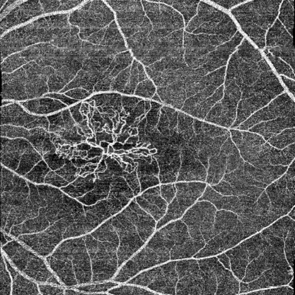
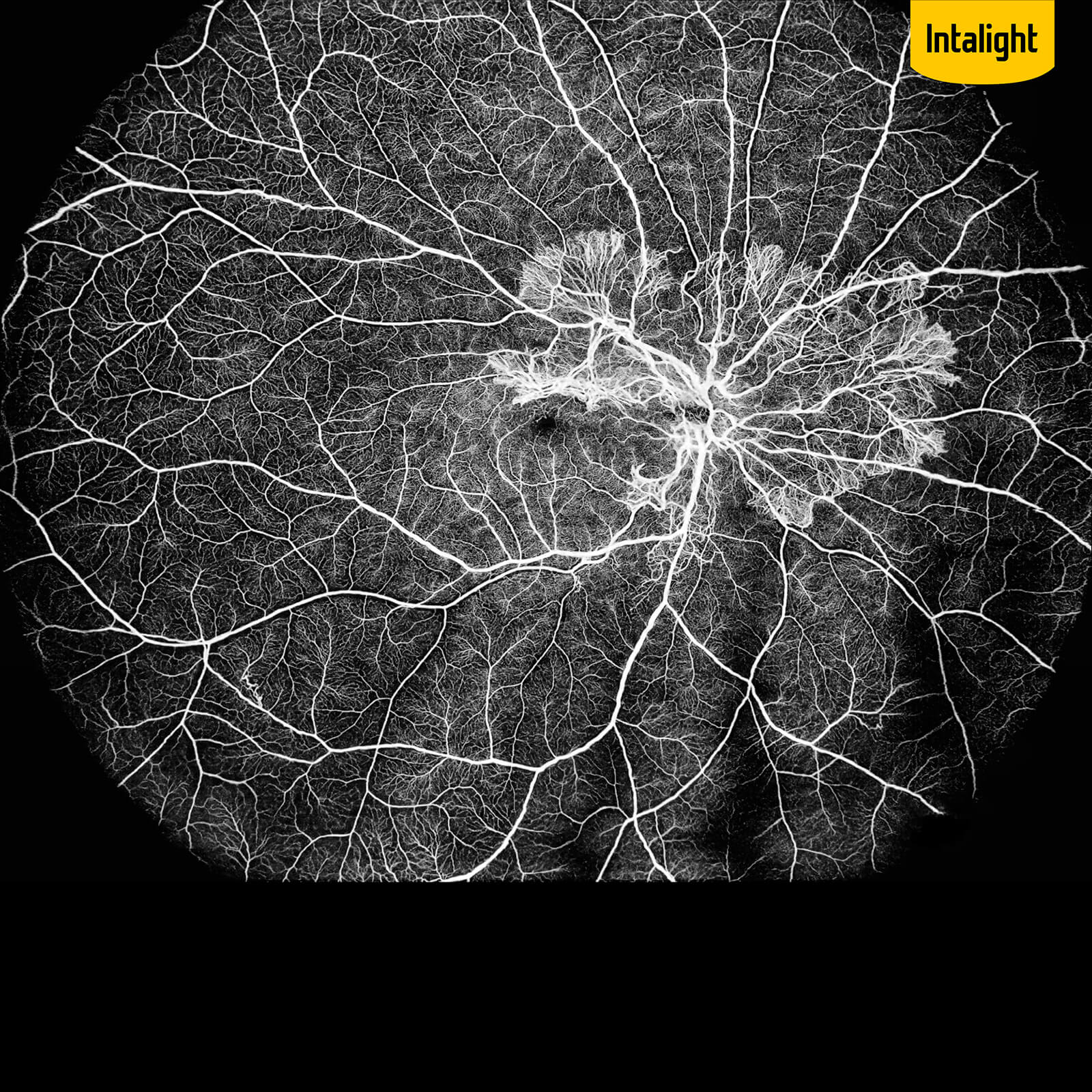
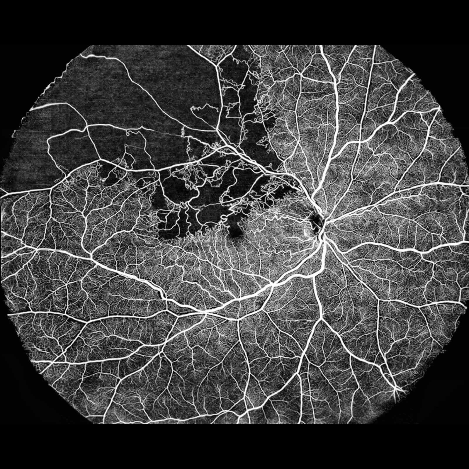
Retinal OCTA

as-oct

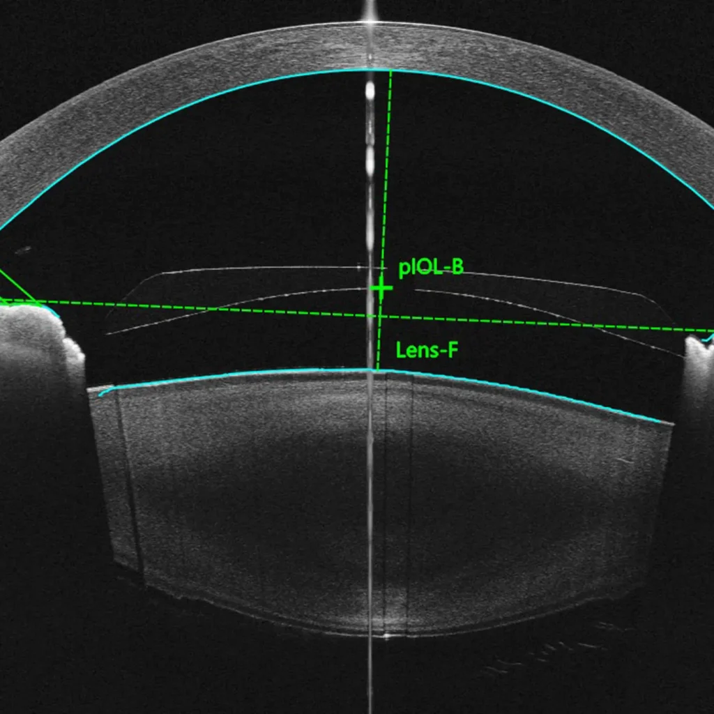
AS- OCT

retinal_oct

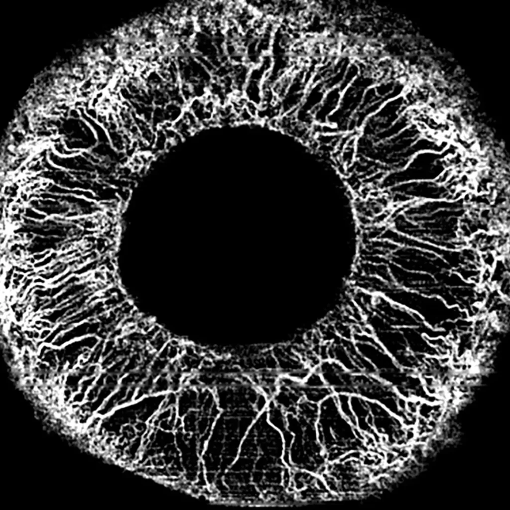
AS- OCTA

retinal_octa

Biometry

Technology

Deep

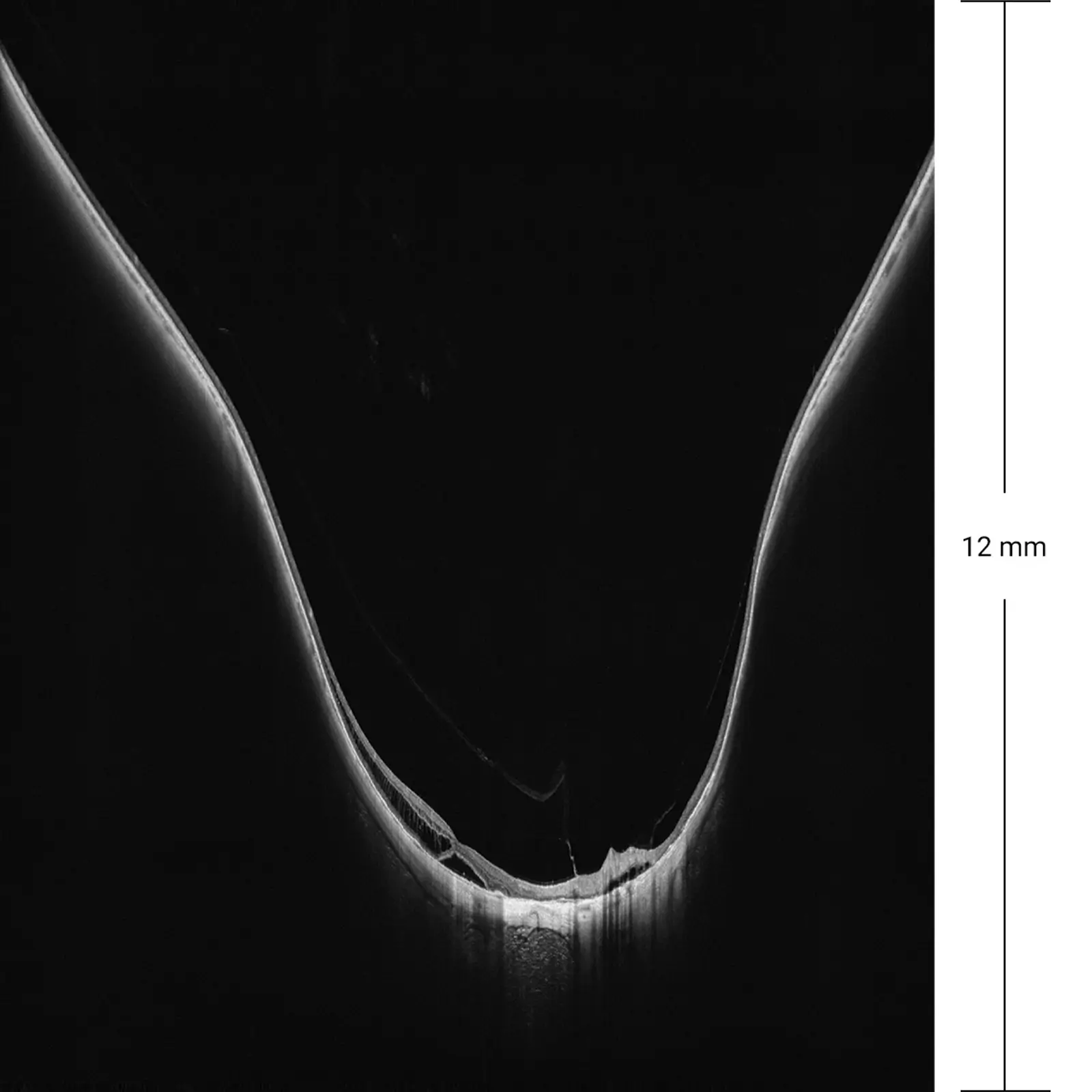
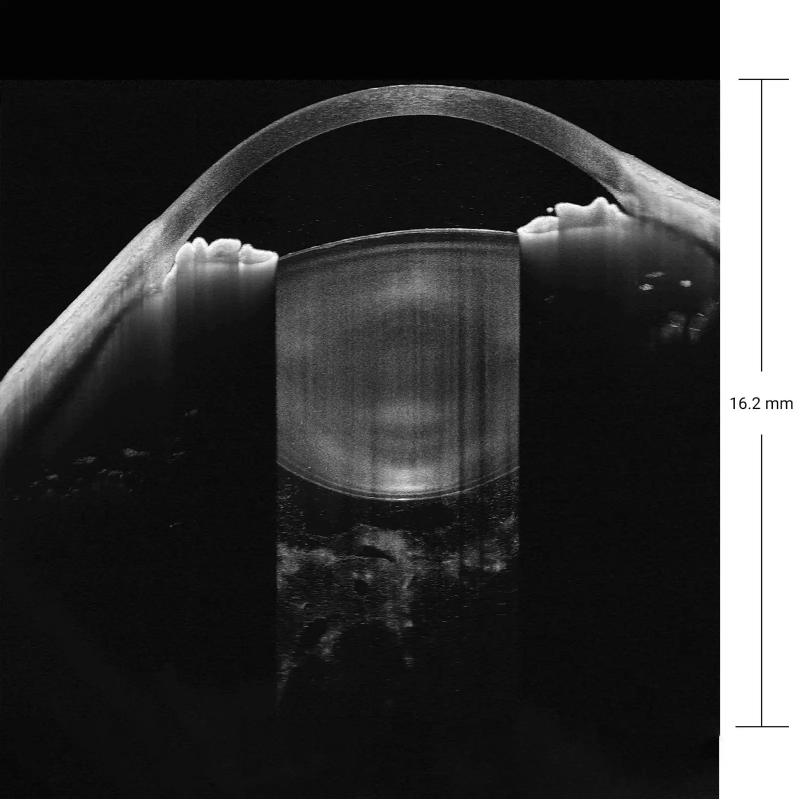
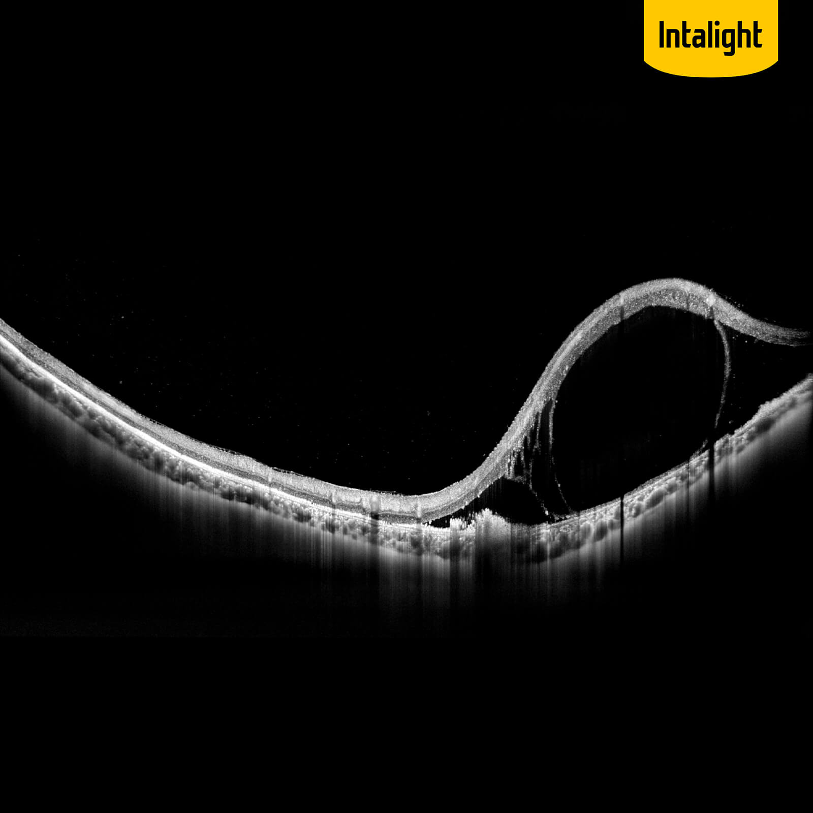
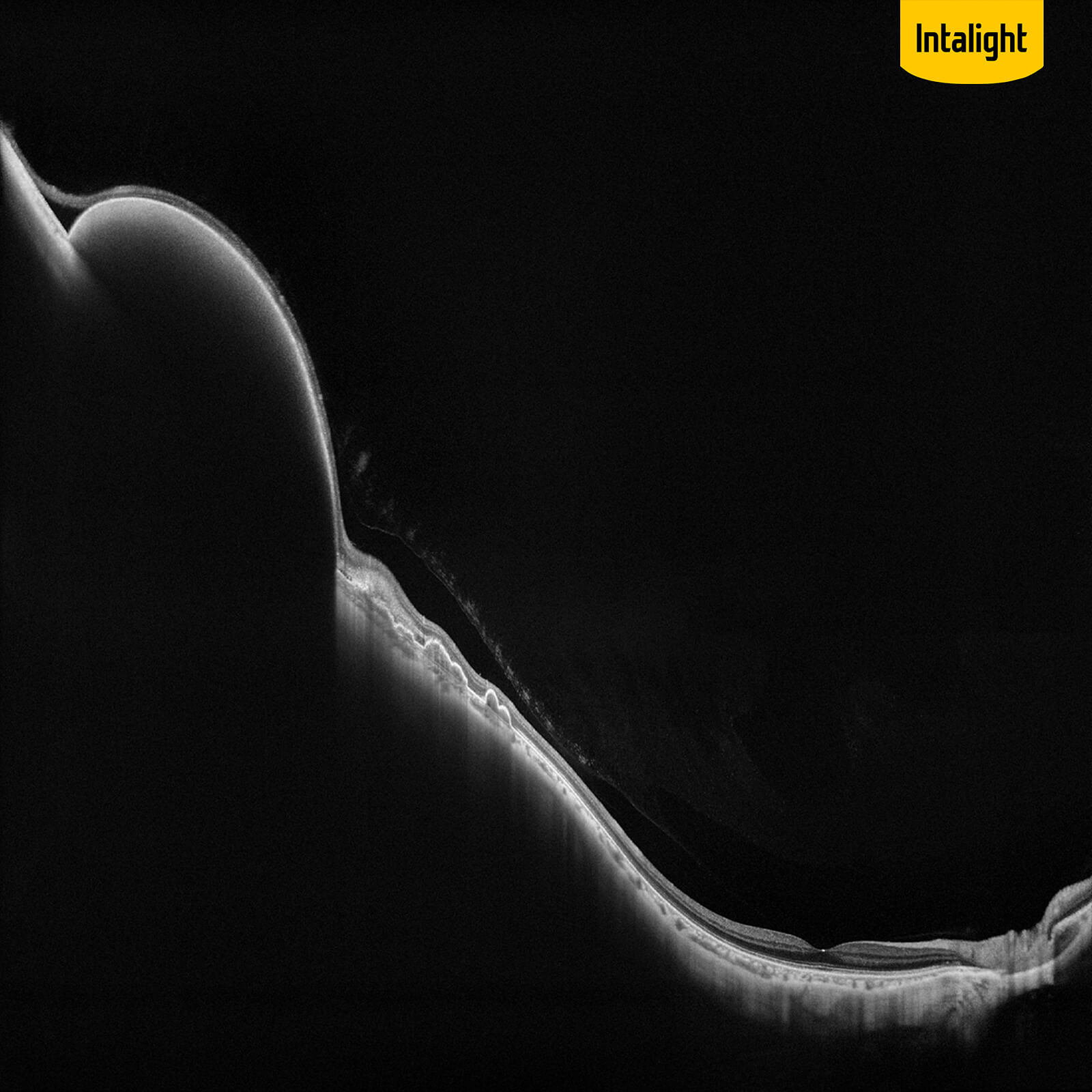
Based on Swept-Source OCT and an proprietary system, DREAM OCT™ can achieve an unprecedented imaging depth of 12 mm (in tissue) for retina imaging and 16.2 mm (in air) for anterior segment.
Rapid
With a scan speed up to 400KHz, DREAM OCT™ is faster than ever before. The high speed is crucial in high resolution OCT Angiography and eliminate the artefact caused by eye motions.

*Product registration varies by country, and scan speeds are available at 400,000/200,000/100,000 A-Scans/s.

Comparison of Scan Speed

DREAM OCT™ up to 400,000 A-Scans/s

High-end SD-OCT

SD-OCT

Extensive

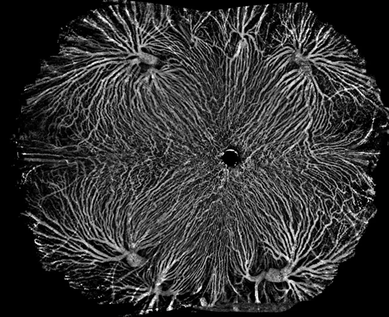
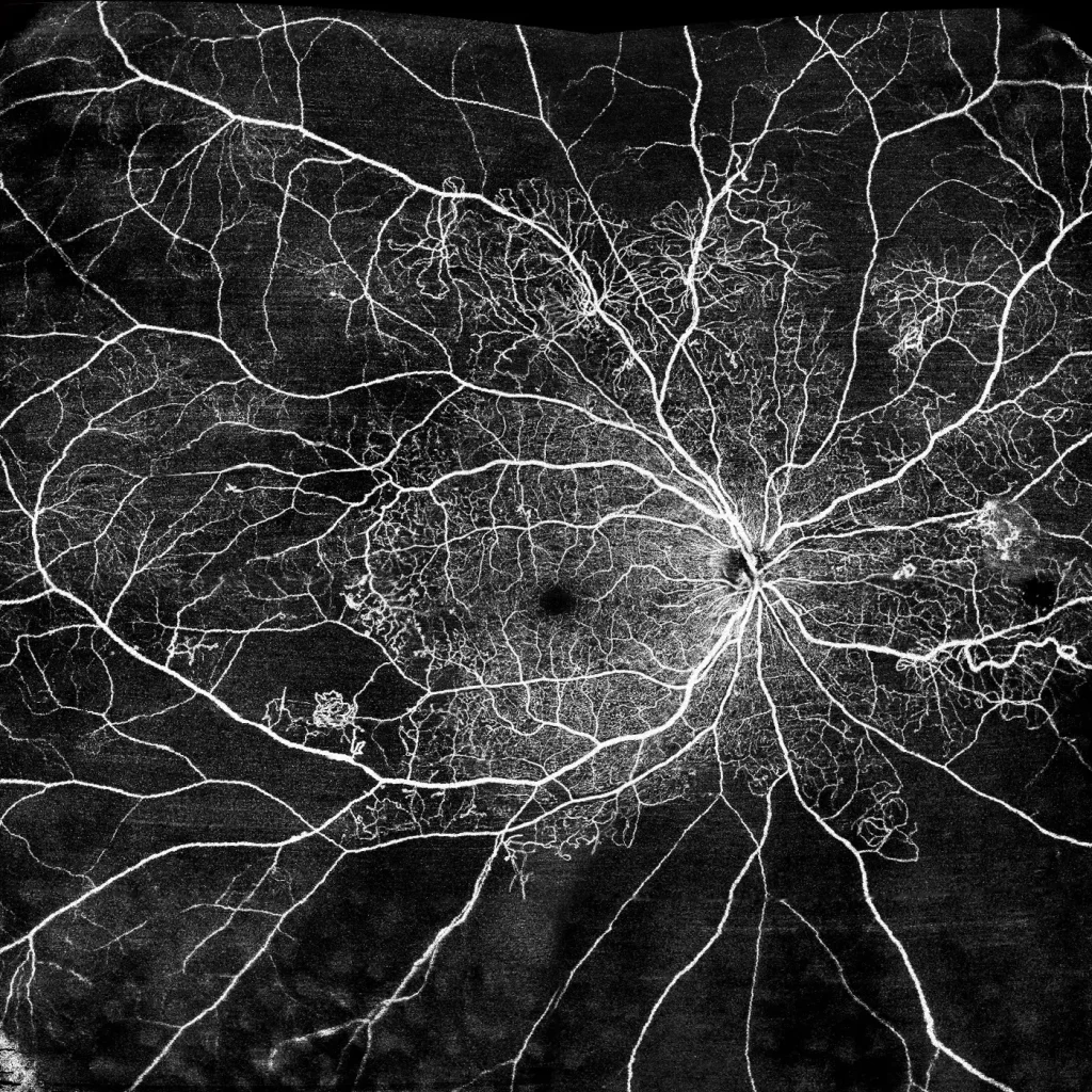
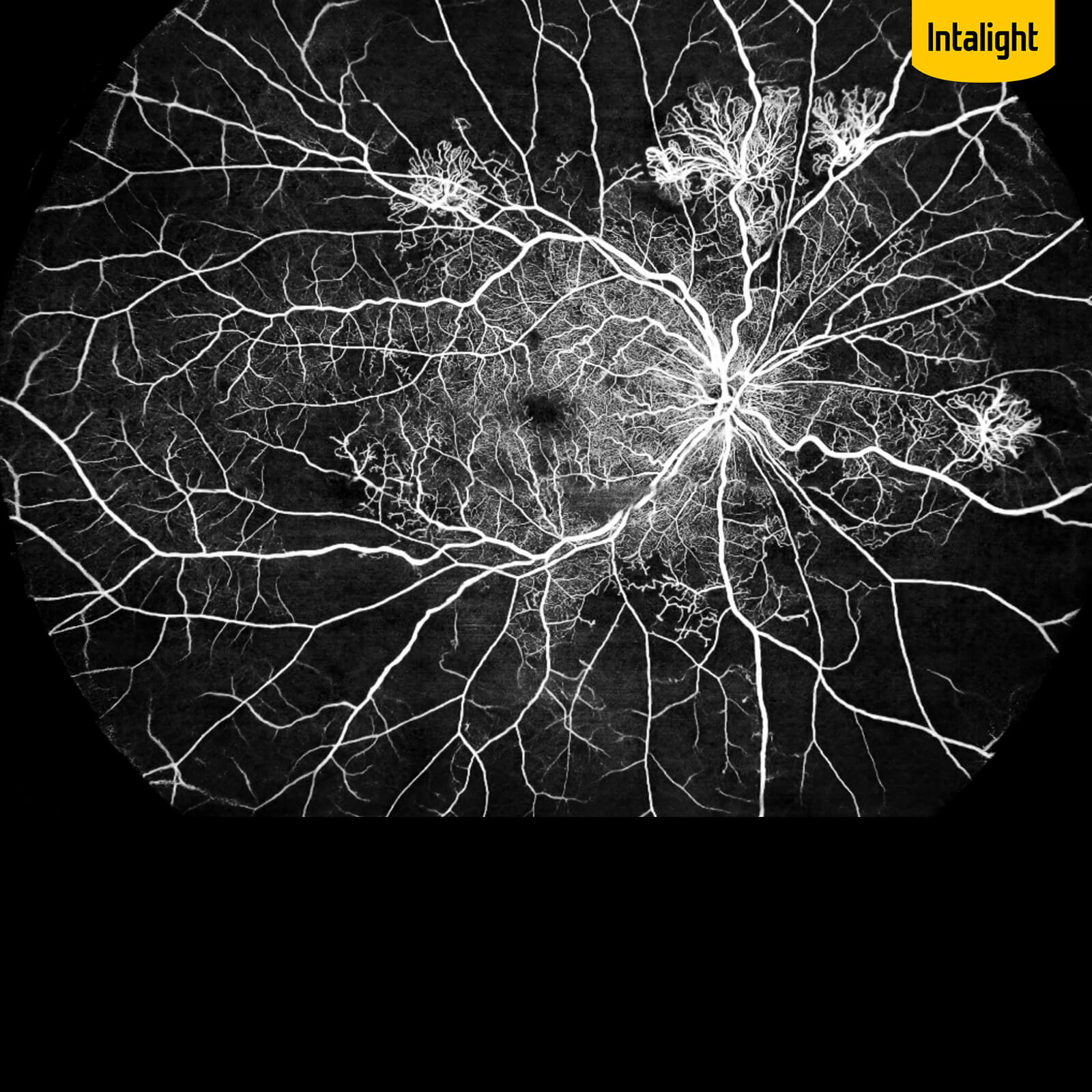
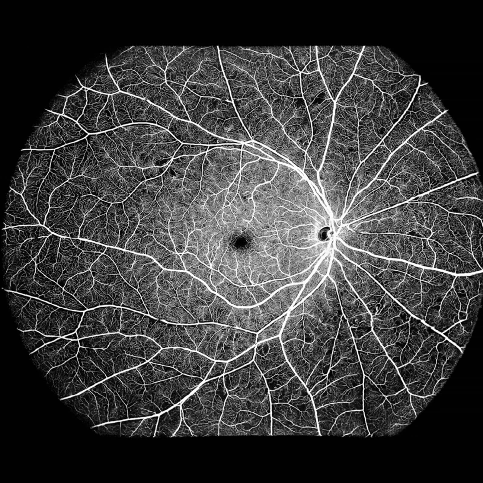
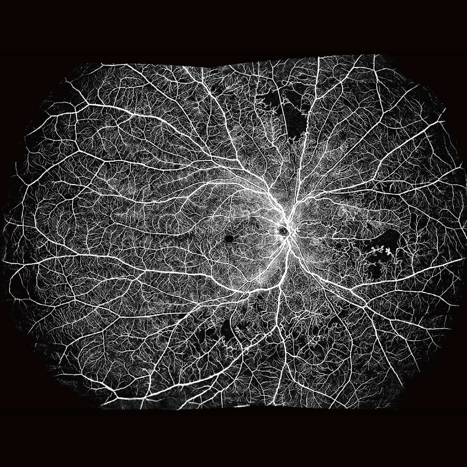
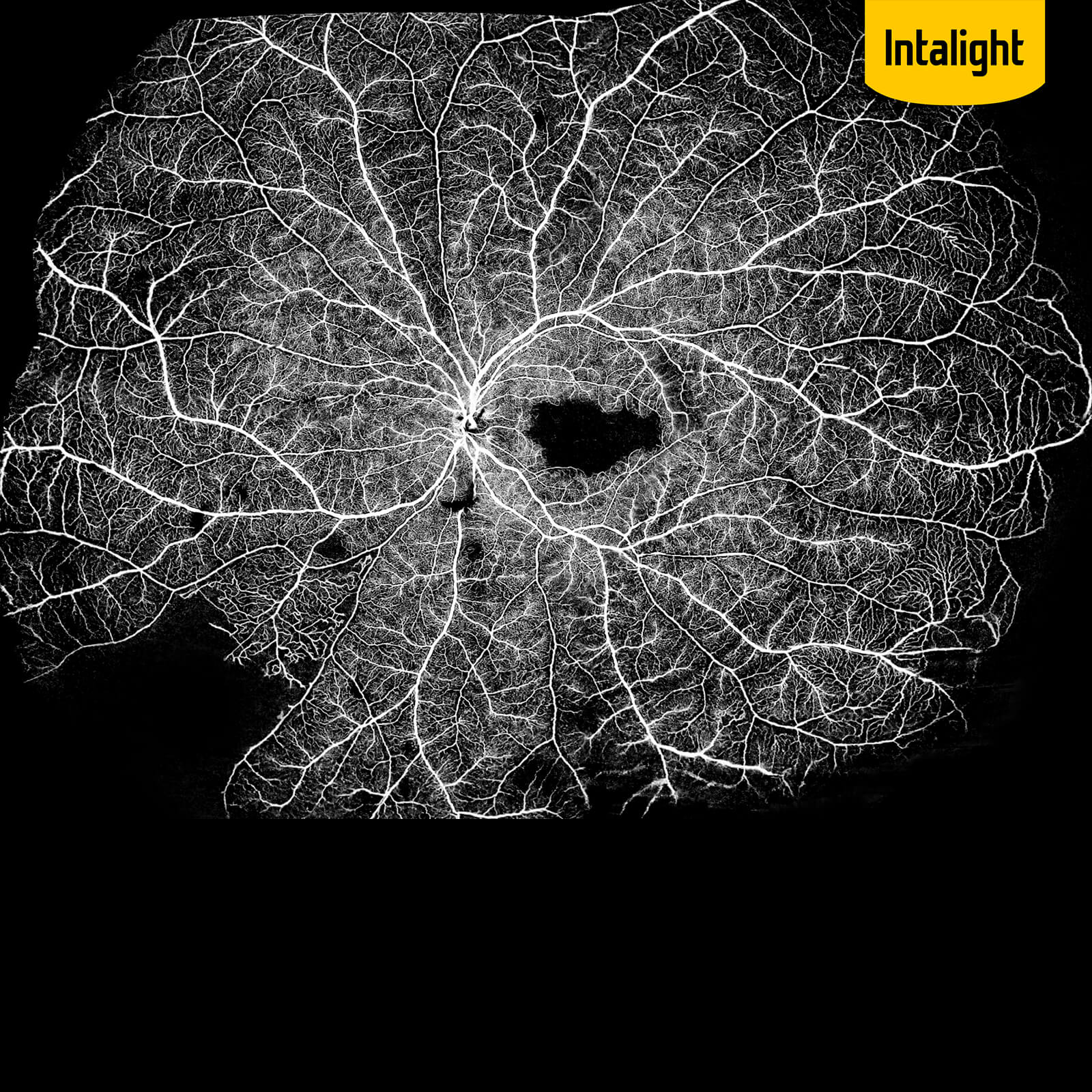
DREAM OCT™ is able to cover an ultrawide field of 26 mm × 21mm (inner angle 130°) on retina with a single scan.
An automatic montage can further extend the range to 200°, going beyond the entire posterior hemisphere and uncovering lesions toward the edge of retinas.

Accurate

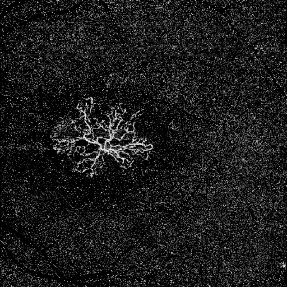
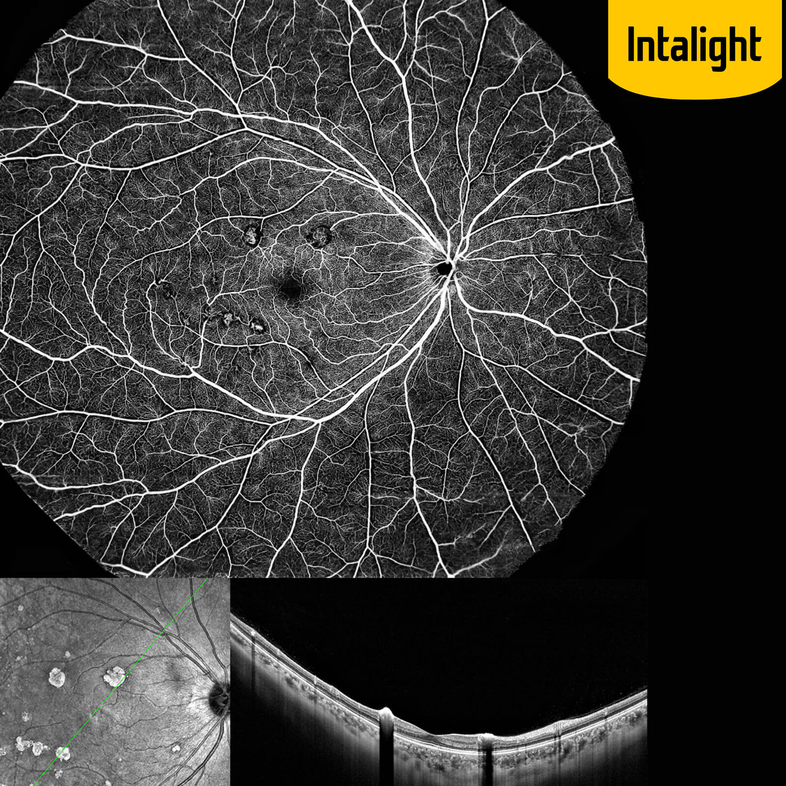
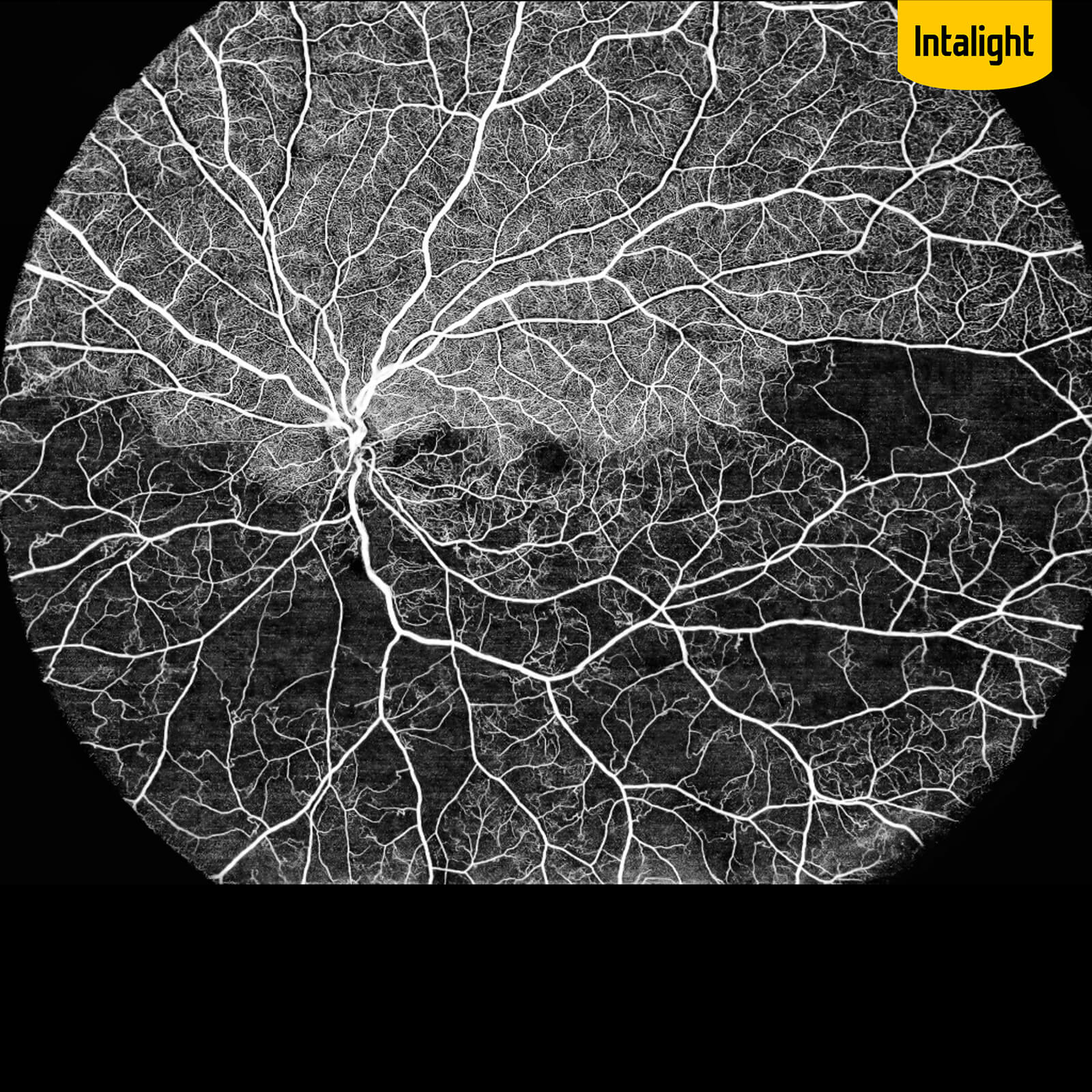
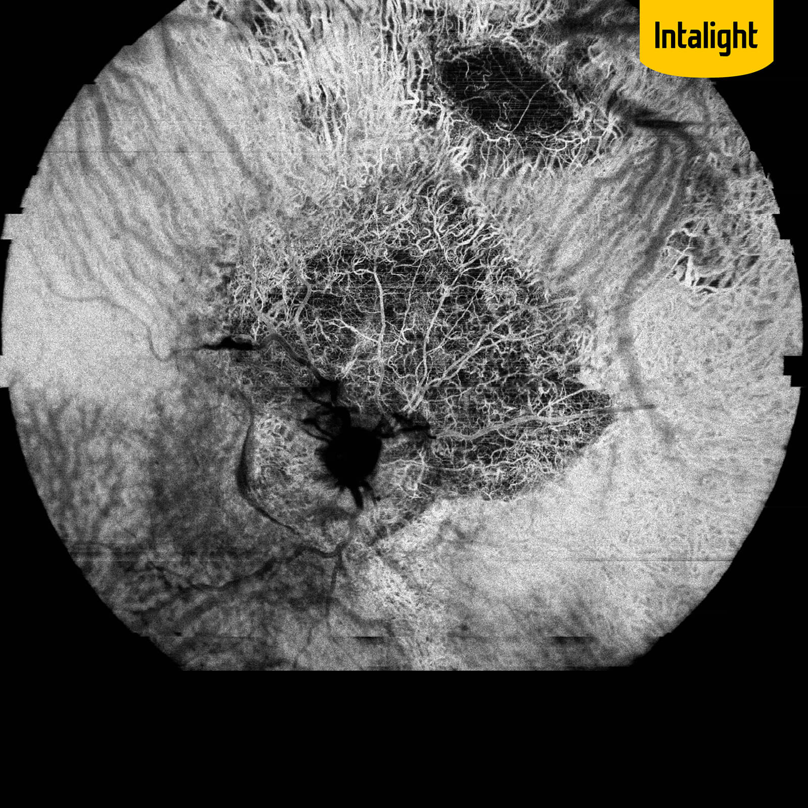
DREAM OCT™ is capable of detecting tiny nascent vascular pathologies while maintaining a low false positive rate. The proprietary algorithm of TrueAngio™ enables a high detection sensitivity of subtle flow signals. 3D Projection Artefact Removal and Deep Layer™ algorithms further help to reduce artefacts to reveal true lesions.
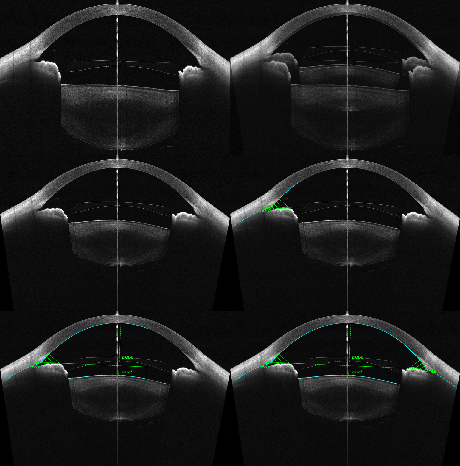
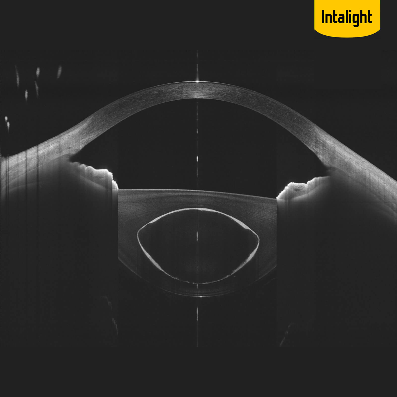
In Anterior Segment imaging and biometric measurement, the raw images undergo sophisticated corrections to account for optical distortion and light refraction. It ensures an accurate and precise quantification of biometric parameters which are crucial for clinical decisions.
Before After

Multimodal

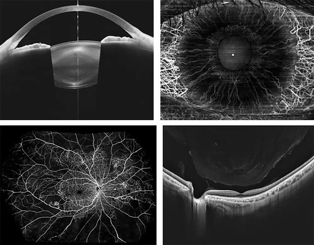
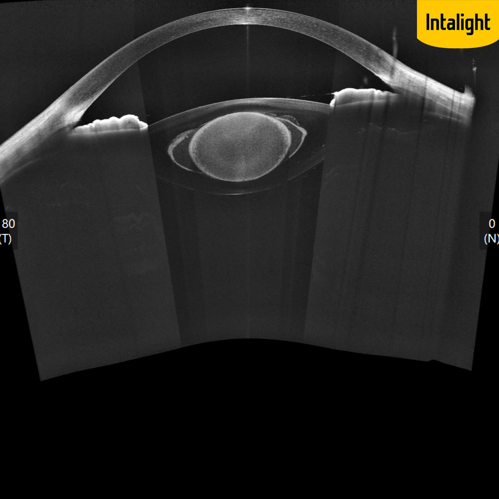
Powered by a state-of-the-art Swept-Source OCT engine, the DREAM OCT™ integrates a full set of imaging modalities including Retinal OCT, Retinal OCTA, AS-OCT, AS-OCTA and visualized biometry for the most challenging clinical and research applications.

The combination of different modalities does not have to compromise each modality’s individual performance. Instead, they work together to collaboratively achieve functions that were impossible before, such as accurate measurement of retina curvature and full eye imaging that reconstructs geometrically accurate 3D representation of eyes.

Application

Retina & Vitreous

Anterior Segment

ACA & ONH

Gallery

Specifications

Item Specification
Corneal Rings24
Measured Points8,640
Camera8 MP
Corneal Coverage10 mm
ImagingColor & Infrared
ConnectivityUSB 3.0

Subscribe Newsletter